Mineral Species
Betekhtinite
Type Locality
No
Composition
(Cu,Fe)21Pb2S15
Crystal System
Orthorhombic
Status at Tsumeb
Confirmed
Abundance
Very rare
Distribution
Second oxidation zone; sulphide ores
Paragenesis
Hypogene
Entry Number
Species; TSNB48
General Notes
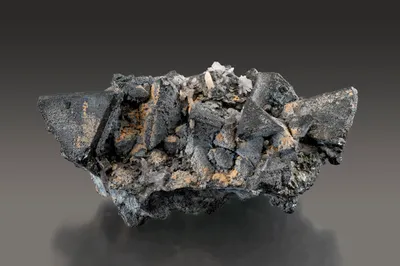
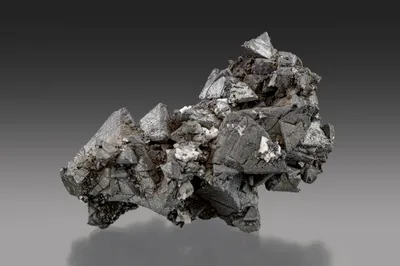
Betekhtinite is generally observable only by reflected light microscopy or electron beam techniques. Specimen material is extremely rare.
The sulphide species originally identified as lautite and ‘pseudo-lautite’ by Geier in unpublished TCL reports was later found to be betekhtinite (Hughes, 1987).
Betekhtinite was first observed at Tsumeb by ore microscopy in the mid-1960s (Bartelke 1976). Strunz and Tennyson (1967) confirmed that betekhtinite had been verified by XRD and electron microscopy and suggested that it had formed as an alteration product of enargite.
Geier and Ottemann (1973) observed that betekhtinite was found almost exclusively between levels 28 and 32, but that a single occurrence was recorded on 24 Level. The betekhtinite was associated with chalcocite, bornite and galena. Based on textural studies, Geier and Ottemann (1973) considered that it formed late in the sulphide paragenesis through the reaction of older chalcocite and bornite with lead-bearing solutions during the formation of younger galena. They further suggested that the relatively limited depth range within which betekhtinite was observed may indicate that very precise physio-chemical conditions were required for its formation.
Electron microprobe analysis of Tsumeb betekhtinite gave the following composition (wt %; average of six microprobe analyses reported by Geier and Ottemann 1973): Cu – 59.2; Pb – 17.5; Fe – 2.8; Ag – 0.8; S – 20.1.
Gebhard (1999) noted the potential for confusion with the acicular habit of chalcocite (and, presumably, furutobeite).
Associated Minerals
bornite; calcite; chalcocite; galena; pyrite; tennantite-(Zn)
Pseudomorphs
Betekhtinite has been reported to form pseudomorphs after chalcocite, although these replacements are microscopic and were observed by reflected light microscopy (rare; Geier and Ottemann 1973).